Gram staining is important for not only categorizing and identifying bacteria but is also used in some medical diagnostic procedures. A study published in the Journal of Clinical Microbiology explored the possibility of using gram staining to diagnose bacterial vaginosis, a pathogenic bacteria. Combining aseptic techniques with gram staining well prepares us to confidently study bacteria in the laboratory and learn more about how bacteria affects the world we live in.
Another important laboratory technique necessary for studying bacteria is staining. The Gram stain process is a staining method commonly used to categorize bacteria and utilizes the unique cellular structure of that bacteria possess. Every bacterium has a cell wall that protects it, provides structure, and prevents lysis in hypotonic environments. Reece et. al. (2011) states that Gram-positive bacteria have a cell wall that is primarily peptidoglycan which retains the violet dye use during the staining process. Contrarily, gram-negative bacteria possess a smaller quantity of peptidoglycan that is layered between the plasma membrane and outer membrane, often with lipopolysaccharide on the exterior. This structure allows the violet dye to be rinsed from the bacterial cell wall leaving a reddish hue visible under the microscope.
After subculturing E. coli we used an inoculating loop to add a small amount of bacteria to a drop of water located on a slide. The slide was then passed through a flame to allow the bacteria to fix to the slide. Crytsal violet was added for about 60 seconds to stain the bacteria before being rinsed thoroughly with water and alcohol to remove any excess dye from the slide. Next, safarin O (another dye) was added in a similar process. A final rinse prepared the slide to be observed under the microscope. The E. coli is gram-negative and appeared pinkish under the microscope.
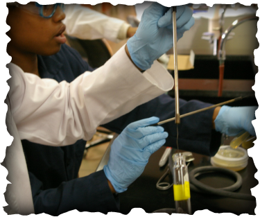
Eden uses an inoculating loop to collect a sample of bacteria from a slant medium
References
Becker, S. (n.d.). Cell Wall Components. Soil Microbiology. Retrieved May 6, 2012, from filebox.vt.edu/users/chagedor/biol_4684/Methods/cellwalls.html
Nugent, R., Krohn, M., & Hillier, S. (1991). Reliability of diagnosing bacterial vaginosis is improved by a standardized method of gram stain
interpretation. Journal of Clinical Microbiology, 29(2), 297-301. Retrieved March 11, 2012, from http://jcm.asm.org/content/29/2/297.s
Reece, J.B., Urry L.A., Cain, M.L., Wasserman, S.A., Minorksy, P.V., & Jackson, R. B. (2011) Campbell Biology. Boston: Benjamin Cummings.
